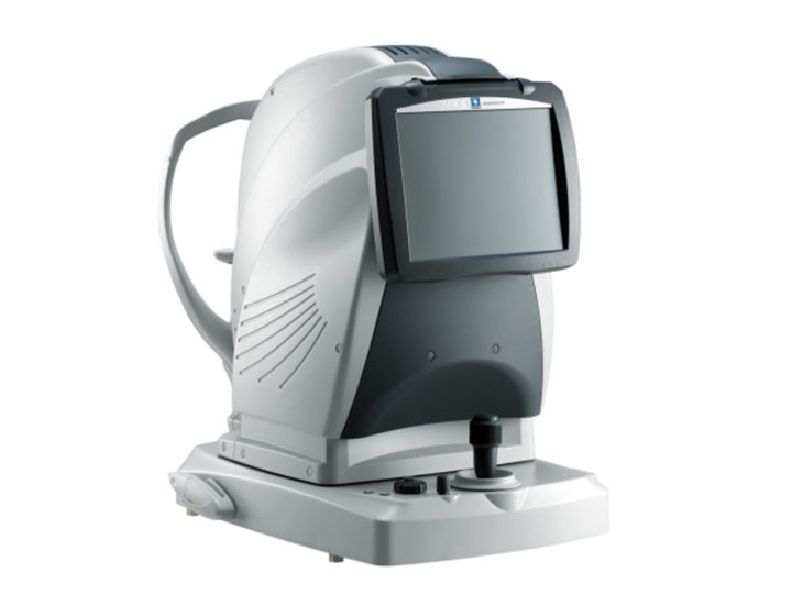

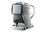
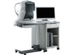
NIDEK MP-3 Micro Perimeter – The Automatic Micro Perimeter With A Non-mydriatic Fundus Camera
- Wide Measurement Range
- Auto Tracking and Auto Alignment
- Fixation Test
- High Resolution Non-mydriatic Fundus Camera
- MP-3 Images of Pre- and Post-treatment Comparison
- Easy to use and simple interface for follow up data review
- Auto tracking and auto alignment
- Robust platform with upgrade options
Further details:
- The MP-3 has a wider range of stimulus intensity, from 0 to 34 dB, compared to the MP-1. The MP-3 measures perimetric threshold values, even for normal eyes. A maximum stimulus luminance of 10,000 asb* allows evaluation of low-sensitivity.
- Auto tracking and auto alignment functions provide more accurate measurements increasing patient and operator comfort and efficiency. These functions allow easy follow-up and reduce variations between examiners, resulting in well-aligned follow up exams.
- The MP-3 can measure fixation and determine the preferred retinal locus, simply by having the patient fixate on a target. Any change in fixation can be compared pre- and post-treatment because the patient’s eye is constantly tracked during microperimetry. This test allows evaluation of fixation in patients with central visual field defects and determines whether fixation improved after treatment.
- The 12-megapixel fundus camera in the MP-3 acquires high resolution images of retinal pathology and allows easy image acquisition.
- Various OCT modalities captured by NIDEK RS-3000 Advance can be registered with microperimetry.