
Including:
- Anterior segment adaptor
- Wide field adaptor
- Remote
- Accessories
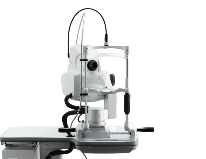
High Definition SLO & OCT Wide Imaging System
All in one device that includes all functions necessary for retinal diagnosis
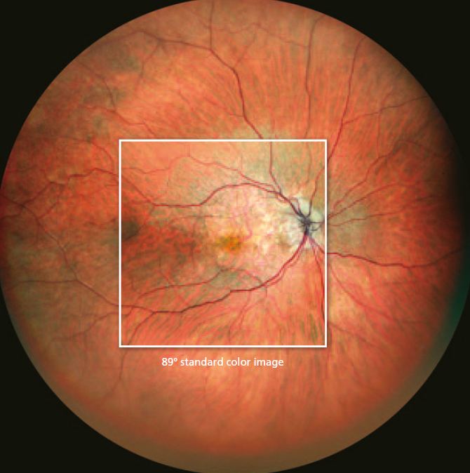
Colour SLO
163º ultra wide field image enables detailed evaluation of of pathologies from the fovea to the extreme periphery.
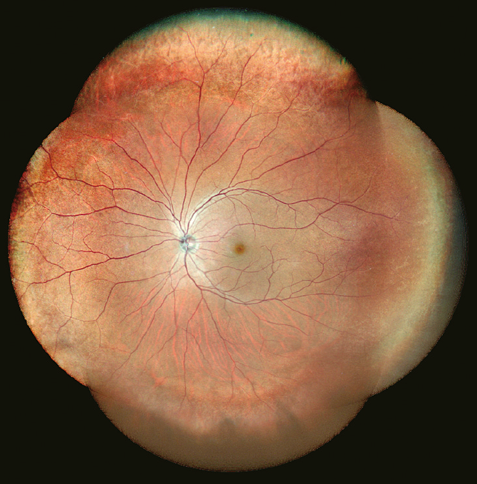
Panorama image composition
With preset fixation points captures details of pathology even in the extreme periphery.
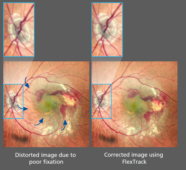
Flex Track Technology
NEW flex track algorithm corrects image distortion due to the instable fixation and enhances averaging quality.
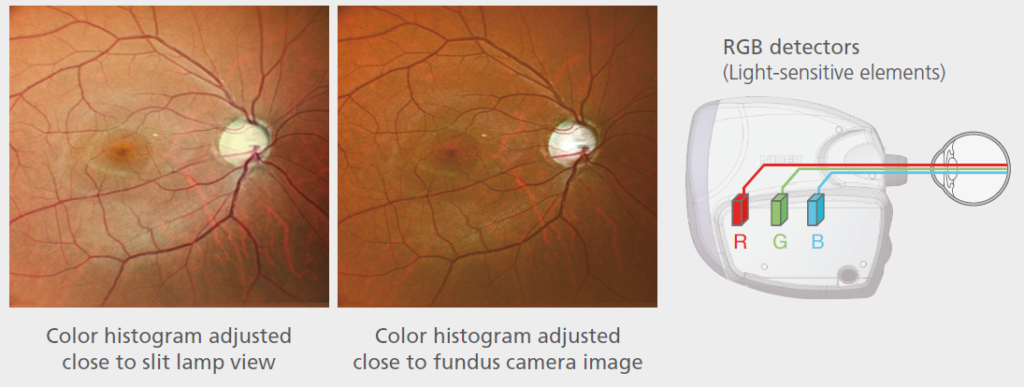
RGB triple detectors
Three seperate RGB detectors simultaneously scan different depths of retina with red, green and blue wavelengths.
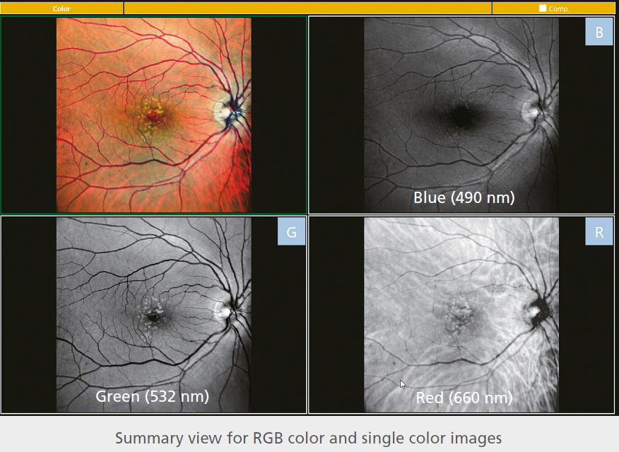
RGB colour & selectable colour display with a single slot
Single colour images in red, green and blue wavelengths can be displayed after colour image acquisition.
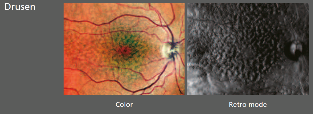
Retro Mode
An unique non-invasive technique for detecting pathologic changes in the choroid
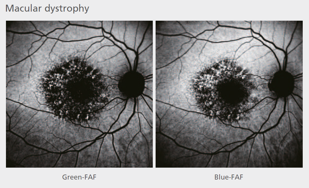
Blue FAF/ Green FAF fundus autofluoresence
FAF imaging is a non-invasive method to evaluate the retinal pigment epithelium (RPE) without contrast dye.
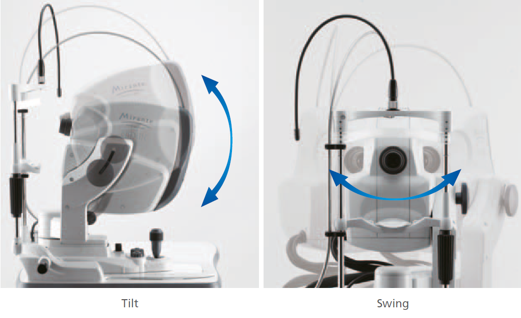
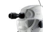
Easy to use functions
Simple interface and easy operation with tilt and swing features
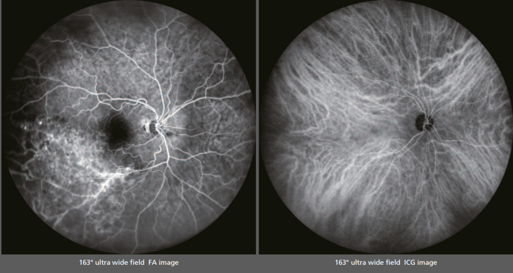
163º ultra wide field FA & ICG images
Ultra wide field imagining is available with the optional wide-field adapter.
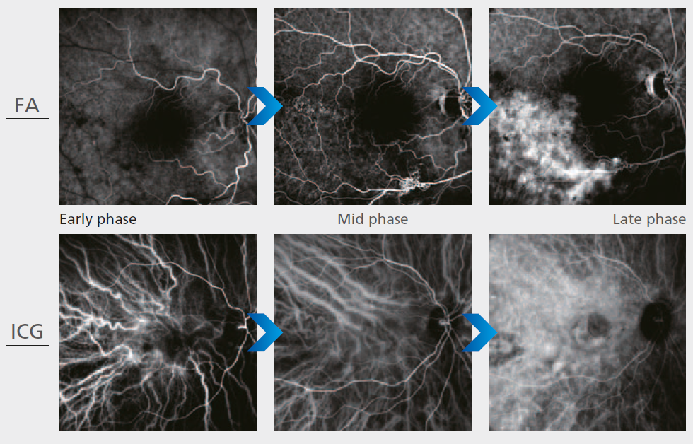
HD dynamics and static angiogram
Auto gain control (AGC) optimises gain level and contrast for early, peak and late phases on angiography.
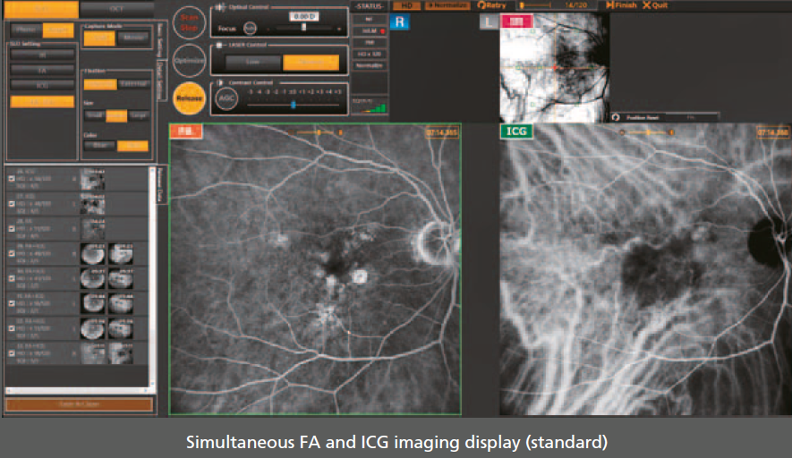
Simultaneous FA and ICG
The Mirante allows simple, simultaneous acquisition of FA and ICG images.
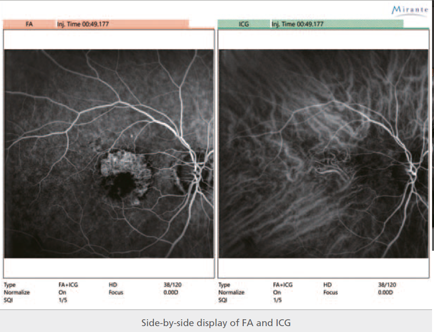
Easy Comparison of FA and ICG
The viewer software can present FA and ICG images side-by-side. Easy comparison is helpful for comprehensive evaluation.
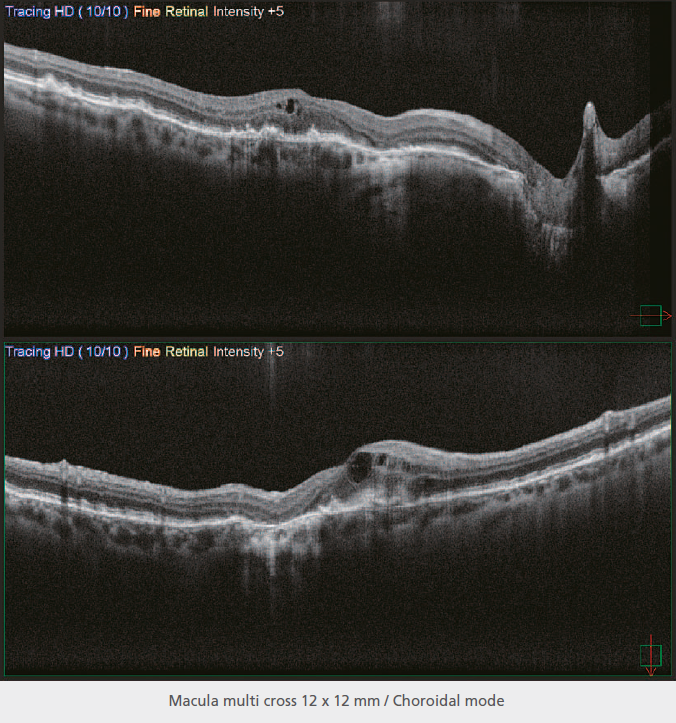
HD wide area OCT
The maximum 16.5 x 12 mm area scan available with the Mirante allows wide area diagnosis including the macula and optic disc in a single shot.
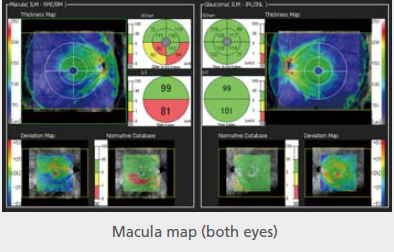
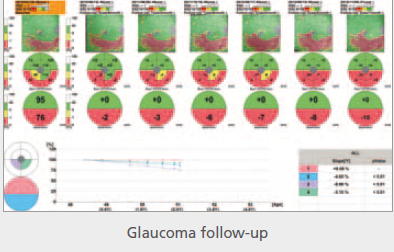
Glaucoma Analysis
The Mirante incorporates 16.5 x 12 mm thickness map which visually presents pathological changes from the central retina to the periphery.
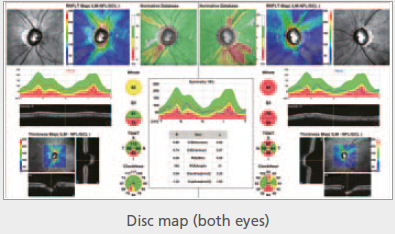
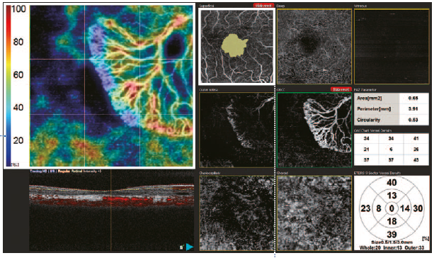
Anglo Scan – Segmentation into multiple tabs
The simple interface provides seven slabs for macula map/ four slabs for the disc map with intuitive functionality and removal of projection artefacts:
– Vessel density map and perfusion density map
– Auto detection of FAZ and shape analysis
– Wide area scan
– Tracing HD plus
– Selectable definition
For full product details on each section please see the product brochure, under related documents.
NIDEK Article – Comparison between two multimodal imaging platforms CLICK HERE