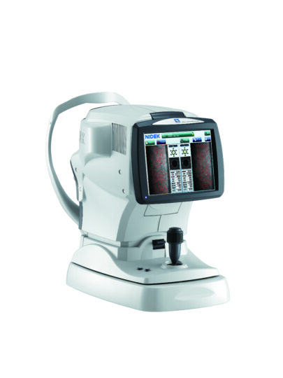
NIDEK CEM-530 Specular Microscope – Combination of Central, Paracentral, & Peripheral Imaging
Multi Area Specular Microscopy
In addition to conventional central and peripheral specular microscopy, the CEM-530 Specular Microscope includes a NIDEK original function that captures paracentral images. The combination of central, paracentral, and peripheral imaging provides a broader, overall view that can be used for detailed morphological and quantitative evaluation of the endothelial layer and individual cells.
Enhanced Usability and Quick Analysis
The 3D auto tracking and auto shot functions result in a user friendly and patient friendly experience. Data analysis within 2 seconds allows efficient patient flow.
Advanced Manual Analysis Functions
Centre Point: Select the approximate centre of a cell. The cells are detected based on the surrounding points. This method is effective for areas where groups of cells are clumped together.
Corner Point: Trace the outlines of the cells to be analysed by selecting the corners of each cell. This method is suitable for detailed identification of the size and dimension of isolated cells.
Pattern Select: Select a hexagonal reference pattern that is similar to the cell size and drag it onto the cell to be analyzed. This method is effective for rough identification of the size and dimension of the cells.
Combination of Auto and Manual Analyses
All three manual analysis methods can be performed on the same image and on auto-analysed images. The versatility of combining automated and manual analysis on the same image allows for better clinical interpretation of the diverse range of pathology in a comprehensive practice.
At Birmingham Optical we also supply a variety of accessories and consumables, which would be relevant to this product. Please visit our store here