
The NIDEK RS-330 now has a software upgrade option.
NIDEK have spent many years perfecting their excellent RS-330 Duo OCT and now they have released new upgrades to the existing device and, excitingly, they have released the Retina Scan DUO II OCT (RSD 2).
The Options Available
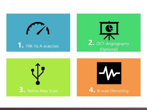
The new software options are:
- New Retina Map Scan
- B-Scan denoising
Retina Map Scan
The Retina Map Scan was introduced by NIDEK to hugely speed up scan capture times for many patients. This rapid screening scan is approximately 55% faster than the current combo scan.
It is also much easier for the patient to follow and it is easier to capture scans. The Retina Map Scan combines the most important elements of the Macula Map, the Disc Map, and the Colour Fundus image (see Figure-1).
All these elements are captured in one rapid single scan per eye, reducing scan capture time and making interpretation much simpler and faster. The Retina Map Scan does not currently use follow up and progression analysis options, but they are soon to arrive. However, Birmingham Optical has devised a new ‘Combo 2’ scan which will allow for progression analysis to take place where it is ongoing and is still faster than the current combo scan.
Figure 1 – The new Retina Map Scan (blue box shows macula analysis and the green box shows glaucoma analysis).
NIDEK RS-330 Duo Software Upgrade Screen
B-Scan Denoising
This incredible upgrade improves the quality of all B-Scan images, even when you scroll of centre on a Macula Map. This makes it much easier to spot subtle lesions, even on Disc Map scans. Best of all, it works retrospectively on all scans.
Figure 2a shows an off-centre B-Scan from a macula map. As we’ve grown to expect, the scan is noisy, grainy, and hard to quickly interpret.
Figure 2a – standard off-centre B-Scan from the Macula Map
NIDEK RS-330 Software Upgrade Scan Image 1
Figure 2b demonstrates the same scan using the denoising algorithm. No new hardware is required; it’s all done by the computer.
Figure 2b – hugely improved B-scan using denoising of the same scan in Figure 2a.
NIDEK RS-330 Duo Software Upgrade Image 2
There are many circumstances where this could make a huge difference to how you manage patients, as you’ll more likely spot subtle lesions. This will only add to your confidence in interpreting OCT results. Even the images on the new Retina Map Scan can be denoised.
Call our team today to find our the difference this will make to your OCT! Call 0808 123 2020
Find out more about the NIDEK Retina Scan Duo 2 OCT, click here